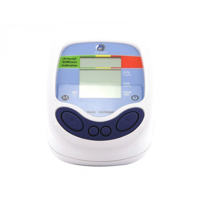
Bio Clip And Pulse Wave Analysis (PWA)
The following report presented as a series of summary statements that are based on the existing published research on Pulse Wave Assessment of Arterial Stiffness. Its aim is to highlight what is known about the reliability of Pulse Wave Analysis as a measure of Arterial Stiffness. At the outset is it worth commenting that the correct reference is to Arterial Stiffness and NOT Arterial Elasticity. They are not the same and Pulse Wave Analysis should not be taken incorrectly to indicate a measure of elasticity. It is also important to note that the assessment of arterial stiffness has been largely conducted by Pulse Wave Volume analysis where carotid artery and femoral artery pulsations are measured and the time delay between the two yields a measure of arterial stiffness. Reflection wave analysis or Pulse Wave Assessment is not the same and its merits must be considered in the context of the information below.
Introduction
Cardiovascular disease (CVD) is the number one cause of death in developed and developing nations of the world, and hypertension is the most important of the multiple risk factors responsible for CVD. Arterial stiffness is the most significant haemodynamic factor contributing to the development and worsening of hypertension Franklin, 2007). Arterial stiffness is recognised as an important and independent risk factor for cardiovascular events (Wang et al, 2008).
Stiffening of the arteries is a common feature of ageing and is exacerbated by a number of disorders such as hypertension, diabetes and renal disease and insulin resistance (Yki-Jarvinen & Westerbacka, 2007). Arterial stiffness of the large elastic conduit arteries is considered a risk marker of vascular ageing (Franklin,2007).
Linkage and polymorphism studies reveal that arterial stiffness in influenced by a first group of genes affecting the renin-angiotensin- aldosterone system, elastic fibre structural components, metalloproteinases, and the Nitric Oxide (NO) pathway. A second group of genes that are involved includes beta-adrenergic receptors, endothelin receptors and inflammatory molecules (Lacolley et al, 2009).
A number of factors are associated with arterial stiffness and may even contribute to it, including endothelial dysfunction, altered vascular smooth muscle cell function, vascular inflammation, and genetic determinants (Wang et al, 2008). Recently, a significant co-relationship was demonstrated between arterial stiffening (assessed by PWV) and inflammation as measured by elevated levels of circulating high sensitive C- reactive protein, tumor necrosis factor-alpha (TNF alpha), and interleukin 6 in essential hypertension (Amar et al, 2005; Mahmud & Freely, 2005).
Epidemiological evidence suggests that insulin resistance and arterial stiffness are interrelated. Insulin acutely decreases stiffness (measured by PWV) in healthy insulin sensitive subjects (Wang et al, 2008). Offspring of diabetic parents have been shown 2 to have carotid artery stiffening in the absence of metabolic or blood pressure alterations (Giannattasio et al, 2008).
There is abundant evidence that arterial stiffness is correlated with abnormalities of lipid and carbohydrate metabolism, which are associated with being overweight and obese. Aortic PWV is associated with visceral adiposity and waist circumference in the elderly. PWV correlated with body mass index from adolescence until old age after full adjustments (Zebekakis et al, 2005).
With a multiple regression study age and albuminuria were independent predictors of aortic PWV (Smith et al, 2005).
Ageing of the arterial system is accompanied by progressive structural changes, consisting of fragmentation and degeneration of elastin, increases in collagen, thickening of the arterial wall, endothelium damage, and progressive dilatation of the arteries (Lakatta, 2003; Safar, 1999). These structural changes in arteries are invariably associated with vascular stiffness as represented by increase in the velocity of the pressure wave as it travels along conduit vessels. As the aorta stiffens, the velocity of the pressure wave increases, and the reflected pressure wave eventually reaches the heart earlier, that is, at the end of systole instead of diastole, causing augmentation of the systolic blood pressure.
The prevention and treatment of cardiovascular disease has focussed on favourably modifying risk factors, such as hypertension, smoking, hyperglycaemia and dyslipidaemia that are known to be detrimental to arterial integrity. However, a significant number of cardiovascular events occur each year in persons who do not qualify for treatment based on these guidelines. It has been found that the addition of other biomarkers such as CRP, BNP (B-type naturetic peptide), NT-proANP (Nterminal pro-atrial naturetic peptide), aldosterone, rennin, fibrinogen, D-dimer, PAI-I (plasminogen activator inhibitor type 1), homocysteine and urinary albumin-tocreatinine ratio, add very little to the classification of risk overall, but greatly increase cost (Wang et al, 2006). In any case, most traditional cardiovascular risk factors alter the structure, properties and function of arterial blood vessels (Hamilton et al, 2007).
It has been suggested that aortic stiffness can occur in the absence of atherosclerosis (Avoilo et al, 1985).
It is recognised that the consequences of risk factors vary significantly between individual patients, and this individual response may be detectable by measuring signs of organ damage or vascular alterations. We may be able to distinguish the beneficial effects of an intervention on an individual’s arterial end organ, rather than simply measuring its effect on intermediate risk markers, such as BP improvements, which do not confer benefits for all individuals.
The Method
Photoplethysmography is inexpensive and unlike tonometry, is operator independent. The only drawback is the damping of the signal as a result of peripheral vasoconstriction. In Japan Photoplethysmography has been widely used by Takazawa 3 et al to assess the effects of vasoactive drugs and characterize vascular ageing. These authors have shown that the second derivative of digital volume pulse (DVP, note this is the same as PWV) may be used to infer changes in the systemic circulation relating to the effects of drugs and ageing. The physical properties determining the characteristics of the DVP remain poorly understood (Chowienczyk et al, 1999).
PWV can be determined from measurements of pulse transit time and the distance travelled by the pulse between the common carotid and femoral arteries. Aortic PWV (carotid to femoral) is a direct measure- the “gold standard” – while PWA and augmentation index are indirect measures of arterial stiffness that also include wave reflection effects (Franklin, 2007).
The central pressure waveform is produced by two major components, a forward travelling wave generated by ventricular ejection and a reflected wave arriving back from the periphery. The reflected wave is influenced by the elastic properties of the entire arterial system, including elastic and muscular arteries. Furthermore, the reflected wave is influenced in part by the magnitude of the incident wave and in part by the balance of vasoconstriction and vasodilatation in the peripheral circulation.
PWV waveform would be expected to be related to the time taken for a pulse pressure wave to pass from the heart to the “site of reflection” and back to the heart. The transit times for pressure waves to pass from the heart to the subclavian artery along the arm to the finger being common for both direct and reflective pressure waves. Calculation of Stiffness Index (SI) is based on the assumption that the subjects height is proportional to the path length of the wave reflection.
The method allows the indirect examination of the structural integrity of both large and small arteries simultaneously allowing the identification of apparently healthy individuals with sub-clinical atherogenesis and premature arteriosclerosis.
Digital pulse volume is sensitive to changes in arterial tone induced by vasoactive drugs and is influenced by ageing and large artery stiffness (Millasseau et al, 2006).
Pulse Wave Assessment provides important information about arterial stiffness. (Wilkinson et al, 1998). It may represent a surrogate endpoint, which may indicate in which patients the traditional CV risk factors translate into real risk.
Reflections from many sites within the vascular tree are likely to contribute to the reflected wave seen at the periphery, resulting in temporal spread of the pressure wave. Several authors suggest that the wave reflection occurs predominantly from the small arteries in the trunk and the lower limbs (Yaginuma et al, 1986; Latson et al, 1988). Reflected waves travel back with approximately the same speed as the forward flowing wave. Propagative models assume that the velocity with which a pulse wave travels along a given artery has a finite value.
Therefore, PWA –is determined by direct and reflected wave pressures. Reflected waves arising mainly from the lower body are delayed relative to the direct wave, and therefore produce and inflection point or second point in the DVP. The phenomenon of wave reflection remains complicated and incompletely understood 4 Changes in peripheral pulse pressure are determined mainly by changes in pressure wave reflection within the trunk and lower body (Millasseau et al, 2000). There is a tendency for the inflection point to be higher in hypertensive subjects. The stiffer the vessel, the faster the pulse wave volume.
Augmentation Index is a term commonly used in PWA and represents the percentage of central pulse pressure attributable to the secondary pressure rise, produced by overlap of the forward and early reflection pressure waves. It depends on variables such as heart rate and vasomotor tone of the arterial system, which can result in considerable variability and thus may limit its use as a surrogate measure of arterial stiffness, especially if a drug under investigation changes the heart rate e.g. beta blockers ( Lacy et al, 2004; Lemogoum et al, 2004).
During dilatation therapy it seems that a reduction in Inflection Point is due mainly to dilatation of small arteries reducing wave reflection from the lower body (Yaginuma, 1989).
Protocol
PWV measured along the aortic or aorto-iliac pathway is felt to be the most clinically relevant ( Nichols & O’Rourke, 1998) since the aorta and its first branches are responsible for most of the pathophysiological effects of arterial stiffness. (Hamilton et al, 2007).
It is important to note that there are several factors that can affect measurement of PWV/PWA and therefore a standardised protocol should be used for all measurements. Some of the potential confounding factors are mentioned below.
In studies of arterial stiffness subjects have been asked to refrain from caffeinecontaining beverages, alcohol and smoking in the 12 hours prior to assessment (as stated above the degree of vasoconstriction will cause dampening of the signal). Subjects were also rested supine for at least 20 minutes in a temperature-controlled environment (24±1 º C). Refraining from talking and sleeping during the reading was also advised. Subjects on anti-hypertensive or anti-anginal medicines were asked to refrain from their morning dose.
Tobacco smoking acutely raises PWV, especially in black subjects, as does consumption of coffee.
Heart rate determines CV and PWC outcomes. There is a direct linear relationship between pulse rate and augmentation index (AI). For every 10 beats per minute increase in heart rate the AI fell by 4%. This finding has been replicated in several studies (Hamilton et al, 2007).
A consensus is necessary on all these issues so that future studies use uniform techniques and analytical approaches.
The Evidence
Measures of arterial stiffness indices are accepted as independent markers of CVD having both prognostic and diagnostic implications. Most methods for assessing arterial stiffness have proven technically difficult or time consuming (Laurent, 2006).
Arterial stiffness measured by PWV analysis has been proven to be a validated and reproducible technique with minimal intra-observer variation (Chowienczyk et al, 1999; Sollinger, 2006).
Stiffness Index (SI) was strongly associated with CVD risk. SI was the best discriminator between low to medium risk and high risk categories when compared to total cholesterol, plasma glucose, systolic blood pressure and waist-hip ratio. In other words SI was better indicator of risk than these other measures. It also had the benefit of discriminating the individuals with known CVD risk factors such as diabetes and hypertension (Gunarathne et al, 2008).
A recent meta-analysis of 17 longitudinal studies to investigate the predicative effect of arterial stiffness measured by PWV of cardiovascular events and all cause mortality showed that aortic stiffness expressed as aortic PWV is a strong predictor of future cardiovascular events and all cause mortality. A total of 15,877 patients were followed for a mean of 7.7 years. The predictive ability of arterial stiffness is higher in those with a higher baseline CV risk (CAD, renal disease, hypertension). An increase in PWV by 1m/s corresponded to an age-, sex-, and risk factor-adjusted risk increase of 14%, 15% and 15% in total CV events, CV mortality and all-cause mortality. CV mortality accounted for only 50-55% of cases of all cause mortality. Vlachopoulos et al, 2010. Pooled relative risk of all cause mortality were higher for high aortic PWV compared with low SI subjects.
It seems therefore that aortic PWV has emerged as an important independent predictor of cardiovascular events (Wang et al, 2008). Its merit over and above other recognised risk factors is that it actually represents a defined change in the target tissue.
One research group has shown that SI was significantly higher in males, older people, those with a history of hypertension, diabetes, hypercholesterolaemia and those with a high waist-hip ratio (but not those with a higher BMI) (Gunarathne et al, 2008).
There is an independent association between waist-hip ratio (WHR) and SI (Gunarathne et al, 2008). Several studies have reported an association of WHR with cardiovascular risk factors such as hypertension, lipid and glucose concentrations (Pouliot et al, 1994). People with raised WHR are also known to have varying degrees of insulin resistance (Westerbacka et al, 2001).
After correction for mean blood pressure (MBP) and age there seems to be no significant influence of smoking status or dyslipidaemia and negligible influence of gender on PWV (RVASC, 2010). As stated above however, there may be an acute affect on PWA measurement from smoking.
Diabetic subjects and subjects treated for hypertension and dyslipidaemia had significantly elevated PWV values, compared with untreated patients, even after correction for age and MBP.
The limitations
Since it is not possible currently to measure directly the mechanical properties of small vessels in vivo, results obtained by model-based analysis of arterial waveforms remain inconclusive and more longterm data about the prognostic capabilities of these techniques are awaited. PWV and PWA do not provide any direct assessment of the mechanical properties of blood vessels (Hamilton et al, 2007).
One of the major drawbacks of the measurement of arterial stiffness is the lack of sufficient reference data for a “normal population”. There are no established cut-offs for stiffness measurements. Efforts at establishing reference values have previously been made, but are usually limited by the size and specificity of the population, yielding results that are only valid in these specific populations.
Very recently (June 2010) there has been a collaborative effort to establish determinants of Pulse Wave Velocity in healthy people and in the presence of cardiovascular risk factors: “establishing normal and reference values” It is called “The Reference Values for Arterial Stiffness Collaboration” (RVASC, 2010). They analysed data from 16,867 subjects from 13 centres across 8 European countries. Of these 11,092 free from overt CVD, non-diabetic, untreated hypertension and not on lipid-lowering drugs. Subjects were categorized by age decade and blood pressure. Subjects other than Caucasian were a small minority. Therefore, we have no idea from this study how it relates to non-Caucasian populations.
They also reiterated that from the different methods to assess arterial stiffness, carotid to femoral PWV has emerged as the gold standard method because of its relative ease in determination as well as its perceived reliability and most because of the large body of evidence demonstrating its association with incident CVD.
The fixed threshold value of 12 m/s proposed in 2007 ESH/ESC hypertension guidelines was based on published epidemiological evidence but could not take into account the multiple factors influencing PWV.
It has been proven that important differences in absolute PWV values exist between methodologies and/or between populations. On the other hand, many risk factors have been shown to influence PWV in small-scale studies and may be confounded by differences in age and blood pressure level. PWV has been shown to have a strong dependence on age and BP (RVASC, 2010).
In the same review it was noted that different alogorithms applied on the same waveforms can lead to differences in measured PWV values of 5-15% (RVASC, 2010). In other words the specific time points chosen for measurement will influence the end result. It is important therefore to standardise these across all research in PWA.
In the same review it was noted that different alogorithms applied on the same waveforms can lead to differences in measured PWV values of 5-15% (RVASC, 2010). In other words the specific time points chosen for measurement will influence the end result. It is important therefore to standardise these across all research in PWA.
Also noted was that differences in path length alone can lead to differences in PWV values of up to 30% (RVASC, 2010).
Evidence for successful intervention on PWV is not yet available.
The RVASC study and others have provided no information about the evolution of PWV over time and as above no outcome data.
Future
The methodology for PWV measurement has not been clearly standardised. Until now there has been a lack of reference of normal values. Recently there has been an attempt to provide normal and reference values for arterial stiffness measured by PWV (The Reference Values for Arterial Stiffness Collaboration, 2010). The results presented are based on a very large cohort of subjects (n = 11,092) with a wide age range. The latter is important since age has uniformly been found to have a strong influence on PWV and values in the elderly may not be applicable to younger subjects. It is only this process of standardisation that will allow for general implementation of this method into clinical practice.
The finding that reflection point PWV response to albuterol depends on the endothelial L-arginine-NO pathway raises the possibility that this response may be used to examine the integrity of this pathway in conditions associated with endothelial dysfunction (Chowienczyk et al, 1999).
Further studies are warranted to assess the utility of SI in clinical practice and to monitor treatment efficacy as well as disease progression.
Perhaps more curiously, it has been found that several diseases seem to be associated with raised PWV for example, rheumatoid arthritis, SLE, overt and subclinical hypothyroidism and metabolic syndrome components (Hamilton et al, 2007). Vascular or Alzheimer’s demetia has also been shown to be associated with higher PWV (Hanon et al, 2005). Rheumatoid arthritis patients were shown to have increased aortic PWV and endothelial dysfunction, as compared to healthy controls ( Maki- Peraja et al, 2006). The elevated aortic PWV was reversible when the inflammation process was reduced by anti-tumor necrosis factor therapy. This suggests that arterial stiffness may be modifiable by the suppression of inflammation.
Tobacco smoking acutely raises PWV, especially in black subjects, as does consumption of coffee. Longterm consumption of coffee is associated with a 13% higher PWV than in persons who do not consume coffee. Moderate alcohol consumption, especially in women, is associated with lower PWV. Acute mental stress and Type A personality behaviour increase PWV.
Helicobacter sero-positivity is also associated with raised PWV in males less then 50 years old. Anti-oxidant status seems to affect PWV too and an inverse assocation of PWV to beta carotene and beta-cryptoxanthin has been reported. Female sex hormones can affect PWV measures. In normal pregnancy, PWV falls in the second trimester, increases in the third and falls after delivery.
Blocking NO synthesis results in acute increase in local arterial stiffness in healthy volunteers (Wang et al, 2008). Increased oxidative stress produced by inflammation inactivates NO, thus inducing endothelial dysfunction. A close relationship has been demonstrated between atherosclerosis and arterial stiffness. Inflammatory mediators can promote leucocyte infiltration and activate vascular smooth muscle cells.
Non-pharmacological interventions shown to reduce arterial stiffness are exercise training, dietary changes and HRT in recent onset menopause (Laurent & Boutouyrie, 2007).
Aortic PWV predicted weight change in a population -based study suggesting that arterial stiffness is sensitive to changes in body weight (Wildman et al, 2005).
Clearly many factors can affect arterial stiffness and what is lacking is intervention data showing that manipulation of these factors can reduce arterial stiffness. A prospective study of the effects of lifestyle changes (e.g. smoking cessation, weight reduction) need to be done as do prospective studies with nitric oxide releasers, antioxidants, anti-inflammatories etc.
In Summary
The assessment of Arterial Stiffness has merit on the basis that it has been shown to be a predictor of not only CVD but also all cause mortality. Furthermore, there appears to be a correlation with a broad range of other diseases. The latter is a curious observation, which is not easy to explain. It may be that stiffness index can be used as general health indicator.
The wealth of evidence for arterial stiffness correlations comes from PWV analysis and not PWA analysis i.e. carotid to femoral PWV has emerged as the gold standard method because of its relative ease in determination as well as its perceived reliability and most because of the large body of evidence demonstrating its association with incident CVD. The same evidence does not exist for PWA and furthermore, there are no published correlations of PWV to PWA results. If there were a direct correlation then this would allow for a correction to be applied.
Only very recently (June 2010) has there been an attempt to publish reference ranges for arterial stiffness. Useful and overdue as this data was, it still has its limitation, as it was based on an almost exclusively Caucasian population so we cannot conclude these reference values relate to the non-Caucasian populous.
It is apparent from the published research that many factors can influence arterial stiffness values. These include heart rate, blood pressure, smoking, caffeine intake, insulin resistance/obesity, alcohol consumption and anti-anginal and anti-hypertensive medications. Some of these are obvious targets for life-style mediated reduction of arterial stiffness but it also raises the issue of standardising the protocol for measurements of arterial stiffness. In order to conduct readings optimally subjects should at least be asked to refrain from smoking, alcohol and caffeine for 12 hours prior to readings. A standardised rest period should also be defined prior to recording to allow for equilibration of heart rate.
There is no published data regarding specific interventional therapy that alters arterial stiffness as measured by PWA. Even though the lifestyle factors named above are known to have an effect on arterial stiffness, there are no studies to show how arterial stiffness may change or indeed the time course of such changes when these lifestyle 9 factors are changed positively. This may prove to be a fruitful area of research since all publication would be novel. In this regard, the effects of various nutritional supplements that may alter nitric oxide production, reduce inflammation, reduce glycation etc remain anecdotal. The question around these would be 1) whether they demonstrate a proven and consistent reduction in arterial stiffness when the latter is assessed correctly and in a controlled manner and whether any effect remains permanently when the supplements are no longer taken. In order to make any claims, this research needs to be done.
Certainly PWV analysis data correlates with CVD outcomes. More interesting perhaps is the association with all cause mortality and with other diseases, some of which almost certainly have an inflammatory component e.g. Rheumatoid Arthritis and Lupus. There seems to be something in this correlation that needs to be explored. Physiological factors that influence arterial stiffness may be related to all cause mortality. Inflammation may be one of endpoints through which these different factors mediate this adverse effect on mortality but there may be others. The main point here that should not be missed is that Arterial Stiffness Assessment may prove to be a useful index of general health and of propensity to disease, There is almost certainly a correlation between factors which increase propensity to disease and the Ageing process itself. It is recognised that arterial stiffness is associated with arterial ageing but a link with ageing in general seems likely since the same factors that age arteries are likely to age cellular processes in general.

