Influence of Peptide Bioregulators on Morphology of Parenchymatous Organs
TABLE OF CONTENTS
Introduction
Chapter 1. Ultrasound diagnosis. Method capacities
Chapter 2. Effect of Peptide Bioregulators on thyroid gland morphology
2.1. Summary on thyroid gland structure and functions
2.2. Thyroid gland disorders
2.3. Current diagnosis methods of thyroid gland disorders
2.4. Thyroid gland ultrasound diagnosis
2.5. Structural changes in the thyroid gland at peptide bioregulators
application
Chapter 3. Effect of peptide bioregulators on the prostatic gland morphology
3.1. Summary on the prostate structure and functions
3.2. Prostate age-related changes
3.3. Prostatic gland disorders
3.4. Current methods of diagnosis of the prostatic gland disorders
3.5. Prostatic gland ultrasound diagnosis
3.6. Structural changes in the prostatic gland at peptide bioregulators
application
Chapter 4. Effect of peptide bioregulators on morphology of other glandular
organs
4.1. Pineal gland (epiphysis)
4.1.1. Summary on the pineal gland structure and functions
4.1.2. Pineal gland disorders
4.1.3. Current diagnostic methods of pineal gland disorders
4.1.4. Structural changes in the pineal gland at peptide bioregulators
application
4.2. Pancreas
4.2.1. Summary on the pancreas structure and functions
4.2.2. Pancreas disorders
4.2.3. Current diagnostic methods of pancreas disorders
4.2.4. Pancreas ultrasound diagnosis
4.2.5. Structural changes in the pancreas at peptide bioregulators
application
4.3. Mammary gland (breast)
4.3.1. Summary on the breast structure and functions
4.3.2. Breast disorders
4.3.3. Current diagnosis methods of breast disorders
4.3.4. Breast ultrasound diagnosis
4.3.5. Structural changes in the breast at peptide
Conclusion
References
INTRODUCTION
Extension of human life span is the most important objective of modern science. With ageing body regulating functions deteriorate and are accompanied by the immunity disorders and development of age-related diseases [1, 2, 7, 21, 33, 35, 37]. The ageing process is characterized with a complex of alterations due to impairments in many body functions, diminution of adaptive capacities, progressive growth of abnormal alterations and death probability. The causes and mechanisms of ageing are not quite clear as yet. A great deal of theories were suggested to explain some or other ageing mechanisms [30, 34]. According to one of them ageing is caused by accumulation of pathologic alterations in the body, and the rate for such an accumulation might be influenced by genetic factors as well as by environmental conditions [11, 12]. An issue of health maintenance and longevity consists mainly in utilization of natural body reserve capacities. Therefore, issues regarding implementation of the very own body biological potential are still of current interest being key objectives for gerontology in general and geriatrics, in particular [47]. One of the priority trends of current research in gerontology is a concept of biological regulation (bioregulation) suggested and proved by V. Kh. Khavinson and V. G. Morozov [25, 26, 27]. The bioregulation as per up-to-date notions proceeds on several levels such as supracellular, intercellular, intracellular and molecular. The bioregulation mechanisms in spite of multilevel hierarchy perform a single task to coordinate processes of biosynthesis, interchanging and reproduction of genetic information. The bioregulation unites all control mechanisms in a pluricellular organism [50].
Multiplicity and complexity of the regulating processes suggest presence of universal mediators for information transmission to a cell. Such mediators are peptide bioregulators that exist in different tissues and reveal a wide range of biological activity. They participate in intercellular interactions by transmitting information recorded through corresponding amino acid sequence from one cell to another [5]. Another and not-the-least objective for gerontology is to pursue for new means of the ageing inhibition and life-span extension (the so-called geroprotectors). Development of new therapeutics based on endogenous physiologic active substances produced by a body is a novel approach for restitution of functions lost with ageing [8].
One of the recent discoveries consists in obtaining from animal organs and tissues of the peptide bioregulators, cytomedins [18]. Firstly the cytomedins were obtained from hypothalamic area of the cerebral cortex, pineal gland, thymus and vascular wall [25, 26]. Later peptides similar in origin and physical-chemical properties but different regarding functional activity were found in other body tissues, as well [23, 41, 42]. At present class of peptide bioregulators enumerates several dozens of known compounds and their number increases constantly. As per chemical composition they are complexes of polypeptides having molecular weight of 1000 to 10000 daltons. After experimental and clinical studies of cytomedins there appeared possibility for preventive and therapeutic application of peptide agents to enhance body resistance to impact of negative environmental factors. These researches admit development of new therapeutic agents containing peptide bioregulators of pineal gland (Epithalamin®), thymus (Thymalin®), prostate (Prostatilen®), brain cortex (Cortexin®), retina (Retinalamin®), etc. [23, 25, 26, 42].
Last years among the agents of bioregulation therapy another new class of therapeutics generally named as cytamins and being biologically active nucleoprotein complexes extracted from animal organs and tissues appeared. The said medicines relate to parapharmaceuticals and can effectively restore altered functions in organs they were extracted from [22, 43].
Normalizing effect of peptide bioregulators on the functions of various organs and tissues was found. One can note one of the mechanisms of <> activity of peptides, namely, participation of cytomedins in regulation of cell proliferation and differentiation, as well as capacity of the self-regulation regarding the number and functional activity of cell elements in population [18, 27]. Therefore, one can suggest that peptide bioregulators might affect tissue morphology restituting their altered structure.
To prove the aforementioned mechanism of action for the peptide agents the clinical study of influence of cytomedins and cytamins on normalization in structure of abnormally altered tissues in parenchymatous organs.
Chapter 1 – ULTRASOUND DIAGNOSIS METHOD CAPACITIES
Among the most topical problems in the modern medicine there stands out the search for such a method of intrinsic organ visualisation that would provide a sufficiently informative, evident, fast and patient-friendly demonstration of the target organs in their normal state and pathologic morphology alterations. We applied one of the radiation diagnosis methods, namely, ultrasound diagnosis (USD).
The USD method presents a number of advantages: it is non-invasive, painless and easily tolerable for the patient, which is very important for any sick person and, especially, for elderly patients. Elimination of ionised irradiation is another USD advantage over the rest of radiation diagnosis techniques. There-upon, USD is virtually unlimited in its application in the patient, i. e. multiple monitoring examinations of the process development are feasible. Providing double-dimension image of the studied organ USD, at the same time, is fast (often used as a screening examination), portable (possible to be used at home, on a trip, etc.), relatively economic (USD costs several times less than, for instance, X-ray examination). USD does not require any special preparation of the patient and has no contraindications, thus allowing immediate examination of various organs [6].
Another reason for using USD is its adequacy for the set objectives. Majority of the target organs are quite accessible for ultrasound. In particular, it refers to the thyroid gland, liver, kidneys, adrenal glands, pancreas, prostate and ovaries. Unfortunately, such structures as the pineal gland, cerebral cortex, vessels are impossible to be assessed by ultrasound. In these cases we used computed (CT) and magnetic resonance tomography (MRT).
We would like to give a brief review of the biophysic basics for obtaining an image by ultrasound.
Ultrasound oscillations appear due to the so-called piezo-effect. In case of the quartz plate deformation, an electric potential appears on its surface. On the other hand, if an electric charge is delivered to the quartz plate its dimensions change. If an alternating voltage with a particular ultrasound frequency is taken to the piezo-element the latter would vibrate with the similar frequency emitting an ultrasound. The piezo-element can interchangeably be both receiver and transmitter of the ultrasound oscillations. One or several suchlike elements mounted in a protective housing along with electric contacts are called an acoustic transformer, transducer or probe. It is an obligatory part for any diagnosis ultrasound device.
Sound spreads in a medium as waves of particular oscillation frequency. In the international system of units (SI) one Hertz (Hz) is taken as a unit of oscillatory motion frequency and equals 1 oscillation per second; 1000 oscillations equal 1 kilohertz (kHz) and 1 000 000 — 1 megahertz (MHz). In medicine audible sound (suggestion, music therapy, etc.), as well as the ultrasound with frequency of 1 to 20 MHz is normally used.
An important role for forming <> width and impulse duration is played by wave-length. The oscillatory motion frequency and the wave-length are of important practical value: the higher the frequency is, the shorter the wave-length is. Application of high-frequency transmitters provides improved exposure of small details in the examined object at little depth, and vice versa.
Acoustic or wave impedance is taken as a characteristic for any elastic medium (including body tissues as well). Changes in the medium elasticity or density alter acoustic impedance. It means that at the medium boundary the sound energy is partially reflected and, furthermore, the larger the difference of acoustic impedance between two media is, the larger the amount of energy would be reflected from the boundary between them.
A boundary between tissues and air is a complete reflector; therefore, lungs or gastrointestinal tract filled with gas cannot be examined by the ultrasonography. Due to the same reason, to provide penetration of the ultrasound wave into the human tissues between the probe surface and skin, one must put a contact medium, i. e. special gel. On the other hand, to conduct intravenous artificial contrasting of heart cavities the so-called <> comprising small carbon dioxide bubbles is applied.
Medical ultrasound diagnosis equipment allows to obtain three types of images, namely, A, B and M. The A-type image is single-dimensional being a single space coordinate along direction of the sound beam spreading. Time is indicated at the X-axis, and the signal amplitude — at the Y-axis. The B-type image is a double-dimensional one of an <> layer obtained at grayscale mode. The less the echo intensity is, the darker the corresponding image part on the screen is, and vice versa. Nowadays the B-type imaging is mainly used in the abdominal sonography. The M-type imaging is based on Doppler’s effect: while a sound source is approaching to an observer, its frequency increases, and at moving away it diminishes. The said principle and imaging type are primary for echocardiography. Basing on the data obtained by M-scanning heart biometric indices (amplitude and speed of motion of cardiac structures, etc.) can be calculated.
Diagnosis ultrasound devices are normally furnished with a set of probes with various radiation frequencies. The probe frequency characteristics significantly alter the quality of the obtained acoustic image. For instance, preferable frequency for examination of the patients with excessive body weight is about 2.5 MHz; organs close to the body surface are better to be insonified using the probe with a frequency of about 7 MHz, and to study the eye structures high-frequency transducers (up to 10 MHz) are used.
Finally, in many cases sonography can and must be considered as preferable, first-choice and major diagnostic method. The given technique often provides all information required at the given clinical situation making unnecessary application of other, more expensive, effort-consuming, aggressive and costly ones [4].
Chapter 2 – EFFECT OF PEPTIDE BIOREGULATORS ON THYROID GLAND MORPHOLOGY
2.1. SUMMARY ON THYROID GLAND STRUCTURE AND FUNCTIONS
Thyroid gland /glandula thyroidea (PNA) [38]/ is a non-pair endocrine gland. Its main function is synthesis and incretion in blood and lymph of hormones regulating tissue growth, development, proliferation, differentiation and body metabolism. This U-shape gland is located in front and at the sides of the trachea and comprises two lobes with different measurements. Generally, the gland right and left lobes are connected with an isthmus. The dimensions of each lobe are: lengthwise — up to 5-8 cm, width — up to 2-4 cm and thickness — up to 1-2.5 cm. During pubescent period the thyroid gland is enlarged, and its size diminishes in the elderly.
The thyroid gland is of histological structure typical for endocrine glands: there are no excretory ducts and every functional unit is closely connected with the vascular system. The thyroid gland structural unit is a follicle, that is a round or slightly oval close vesicle with the wall covered with secretory (follicular) epithelium, i. e. thyrocyte. Thyrocytes produce thyroid hormones (thyroxin — T4 and triiodothyronine — T3), as well as thyreoglobulin. The latter is the main colloid constituent filling the follicular space. Simultaneously the thyroid gland produces thyreoalbumin. Normal ratio of thyreoglobulin to thyreoalbumin is about 9 : 1. In disorders associated with thyroid gland parenchyma proliferation, its goitrous transformation and adenoma development thyreoalbumin production increases, and the latter, in case of thyroid malignancy, might even excess thyreoglobulin synthesis.
Beside the follicles, in the thyroid gland interfollicular (extra-follicular) islands formed by cells with a typical thyrocyte-like structure are defined. Interfollicular islands are of importance for regeneration of the thyroid gland if the latter considerably impaired and entire follicles are destroyed.
Hypophysial thyrotropic hormone is considered to be the specific thyroid gland stimulator. In its turn, thyrotropic hypophysial function is activated by thyroliberin produced in the hypothalamus. Therefore, hypothalamic impairment causes similar decline in the thyroid gland functioning as hypophysectomy. This can be defined as transadenohypophysial regulation.
In its turn, the thyroid hormones inhibit the hypophysial thyrotropic function, i. e. the relationship between the functional thyroid gland activity and the hypophysial thyrotropic function intensity is a negative back-feed system that keeps fluctuations of the thyroid gland activity within the normal physiologic bounds.
The thyroid hormones are necessary for normal functioning of the central nervous system. Content of the thyroid hormones in children at the age of 1 to 15 years does not change considerably. However, with ageing the thyroxin-binding globulin and thyroxin levels decrease and triiodothyronine content increases.
The thyroid gland functional activity remains stable for a long time. Only in the elderly people atrophic alterations in the gland parenchyma are observed. They are associated with inconsiderable decrease of basal metabolism though there are signs of the thyroid gland activity growth that can be considered as compensatory reaction counteracting oxidative processes exacerbation in the tissues of a senescent organism [48].
2.2. THYROID GLAND DISORDERS
Both birth defects and alterations in the thyroid gland are rare. Primarily, the thyroid gland disorders are accompanied by signs of increased (thyrotoxicosis) or inhibited (hypothyrosis) function. However, some disorders of the thyroid gland function can have no clinical manifestation (euthyroid state).
The most wide-spread thyroid gland disorder is endemic goiter found in geographic regions with the shortage of natural iodine supply. In the course of the disease the thyroid gland enlarges though, in the majority of cases, without alteration in its function. Its hyperfunctioning bringing forth abnormal metabolism and development of various disturbances in different organs and systems is called <>. The following types are distinguished: diffuse, nodular and mixed toxic goiter. Diminished activity of the thyroid gland (hypothyrosis) emerges as a result of impairment in the gland, hypophysis or hypothalamus. The following types of the thyroid gland inflammatory disorders (thyroidites) can be distinguished: acute, subacute and chronic.
The thyroid gland tumours are often found along with enhanced hypophysis function that causes proliferation of the thyroid epithelium. The hypophysis thyrotropic function can be stimulated by alimentary iodine shortage, antithyroid agents, ionised radiation, hormonal disorders. There are benign and malignant tumours. Among the benign formations adenomas reveal the greatest incidence (16% of all nodular thyroid swellings). They can be single or multiple (multisite nodular goiter). As per the histologic structure, the following adenoma types are defined: trabecular (embryonal), tubular (fetal), microfollicular and macrofollicular (colloid). Still, fibromas, tetratomas, paragangliomas, hemangiomas, lipomas and miomas are rarely registered. The first place among malignant thyroid tumours (90% of all neoplasms) is taken by cancer. Such non-epithelial tumours as sarcoma, malignant lymphoma, hemangoendothelioma, malignant teratoma are rare in the thyroid gland.
2.3. CURRENT DIAGNOSIS METHODS OF THYROID GLAND DISORDERS
Methods of examination of the patients with thyroid gland disorders include clinical examination, methods for evaluation of the gland function and structure. The clinical examination comprises case taking and objective data (assessment of skin, subcutaneous fat, hair, nervousmuscular and cardiovascular systems, gastrointestinal tract). The thyroid function is estimated by indirect and specific methods. The indirect methods (examination of basal, fat and protein metabolism, state of nervousmuscular and cardiovascular systems) are based on investigation of the organism physiologic functions influenced by the thyroid hormones. Indices obtained by these methods are not specific to the thyroid gland disorders as other diseases might cause similar alterations. The specific diagnosis methods for the thyroid gland functional state include blood tests for the thyroid hormones and iodine metabolism.
The methods for the structure assessment are based mainly on radiation examination (conventional roentgenography, radionuclide examination, sonography, CT and MRT) and needle biopsy. Below the method of ultrasound diagnosis for the thyroid gland disorders is described.
2.4. THYROID GLAND ULTRASOUND DIAGNOSIS
The thyroid gland USD provides highly accurate assessment of the gland position, form and dimensions, structure, surface profile, state of surrounding tissues. It should also be stated that nowadays the sonography appears to be the most optimal diagnosis method allowing fast, informative and painless evaluation of the thyroid gland morphology [49].
To conduct the most effective sonographic examination it is desirable to use high-frequency probes of 5 to 7.5 MHz giving resolution of 3 —1 mm. During the examination the patient is in horizontal position on his/her back, with the head slightly inclined backwards. Such position is comfortable for the patient and does not cause any negative effect.
The normal thyroid gland at transversal scanning looks as a homogenous U-shape formation with medium echogeneity and fine-grained structure covering the trachea. Outside the side lobes there are round echo-negative zones — vessels: common carotids and jugular veins. In front of the gland there are hypoechogenic stripe-like structures — neck muscles. Inside each lobe there is a non-homogenous oval formation — the trachea (Fig. 1).
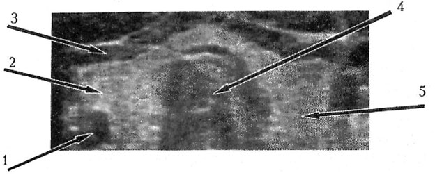
Fig. 1. Transversal sonogram of the normal thyroid gland: 1 — neck vessels, 2 — right gland lobe, 3 — muscles, 4 — trachea, 5 — left gland lobe
USD of the normal gland gives the following dimensions: width of each lobes from the tracheal wall to outward gland border — up to 2-2.5 cm; the lobe length (front-back dimension) — of about 2 cm, and the maximal isthmic length is of about 0.6 cm. At the examination it is advisable to scan in multiple plans providing more detailed examination and structure assessment from all angles (sections) of the gland. The gland lobe length at longitudinal section can reach 5-6 cm.
Pathologic alterations in the gland at the sonography are exhibited with dimensional transformation (changes in the size and form) and local or diffuse echostructure rebuilding.
Changes in the size and form without any structural alterations are mostly found in the patients with endemic goiter. The gland is evenly enlarged owing to both lobes of the isthmus with a form more resembling a large oval.
Changes in the size and form with any structural alterations appear in case of diffuse goiter with or without nodes. If there are no nodes, along with enlargement one can note non-homogeneity of the sonographic structure in its parenchyma — normal tissue sites alternates with hyperechogenic inclusions, zones of rarefaction (diminished sonographic density).
Local parenchymatous reconstitution of the thyroid gland (with or without dimensional changes) is determined mainly with benign (adenomas, cysts) and malignant (cancer) neoplasms. Various adenomas (nodular goiter) are oval or round formations with distinct smooth profile. Inner structure of the gland is mostly homogenous, its sonographic density is decreased but, however, the latter can be equal to surrounding tissues or, more seldom, have increased echogeneity. A characteristic feature consists in the presence of hypoechogenic rim (<>) of about I-2 mm width that is an image of a well-formed capsule, edged out lobes, blood and lymph vessels. Heterogeneity of the node inner structure is caused by degenerative processes as cyst-like cavities of irregular shape or presence of denser wall inclusions in the echo-negative node. Sometimes little calcified formations of high echogeneity can be found in the adenomatous node.
Thyroid gland cyst is a echonegative formation of regular round or oval shape with distinct and even profile. Dorsal ultrasound enhancement can be noted behind the cyst. Often one can find in the gland parenchyma little (2-5 mm in diameter) echonegative cyst-like formations with very smooth and distinct profile that, according to some researchers [16], are centers of local follicular degeneration (focal hyperplasia, colloid degeneration). The said formations are likely to indicate signs of dysthyrosis.
Thyroid gland malignant tumours contribute a little to the morbidity structure and, generally, are rather rare. The highest oncologic alarm is related to solid and mixed solitary nodes. We should also indicate that nowadays there are no exact and reliable sonographic criteria for differentiation of benign and malignant thyroid gland formations. A range of important sonographic signs is somewhat incidental to both types of neoplasms. In some cases malignant tumours can fully reproduce the picture obtained for benign adenoma. Therefore, the sonographic data mostly allow to get a view on the anatomic structure only rather than on the impairment characteristics.
Reconstitution of the thyroid gland sonographic structure is observed also at chronic inflammations, thyroiditis. The most illustrative picture presents sites of irregular form, different sizes, diminished sonographic density and rarefactions against, the unchanged tissue background. The glandular parenchyma appear to be <>. Some researchers consider the hypoechogenic sites as manifestations of the gland stroma edema and lymphoplasmocytic infiltration [16]. Thus, USD as a very effective diagnostic method for external assessment of the thyroid gland is recommended for application at the first stage of examination in the patients with such disorders.
2.5. STRUCTURAL CHANGES IN THE THYROID GLAND AT PEPTIDE BIOREGULATORS APPLICATION
Our study objective was to assess the influence of peptide bioregulator. The complex included immunomodulating agent Thymalin, neuroendocrine system regulator Epithalamin and organotropic agent for the thyroid gland Thyramin on morphologic structure of the thyroid gland parenchyma in case of organic alterations.
Thyramin is a medicine of cytamin class used as a biologically active supplement. It was obtained from the cattle and pig thyroid gland. It comprises nucleoprotein complexes selectively affecting cells of the thyroid gland tissue.
There were examined and monitored 46 subjects at the age of 17 to 72 years with some thyroid gland disorders. Table 1 represents characteristics of the examined subjects and disorder type. Taking into account that in some subjects several types of the gland disorders occurred simultaneously, the total number of revealed nosologies exceeded the number of the patients and amounted to 63 cases.
Table 1
Characteristics of the examined subjects with various abnormal alterations in the thyroid gland
|
Disorder type (nosologic forms) |
Number of cases |
Male |
Female |
Age (years) |
Mean age (X±m) |
| Focal colloid degeneration |
26 |
I0 |
16 |
22-62 |
41.7±0.3 |
| Adenomatous nodes |
20 |
12 |
8 |
43-72 |
57.5±0.3 |
| Chronic thyroidites |
9 |
3 |
6 |
46-67 |
56.5±0.2 |
| Cysts |
8 |
4 |
4 |
33-41 |
37.4±0.1 |
| Total: |
63 |
29 |
34 |
22-72 |
— |
The data given in Table 1 indicate that mostly in our study we noted signs of focal colloid degeneration (focal hyperplasia). The second frequency position is occupied by benign adenomas. The chronic thyroid gland inflammation and the cysts were relatively seldom.
Dysthyrosis (i. e. colloid degeneration of the thyroid gland) were equally found both in the younger patients and in the middle-aged subjects. The gland adenoma was revealed mainly in the middle-age and elderly subjects. Ratio of males to females was approximately equal with little prevalence of females. Multiple signs of disorders were noted in 17 subjects (37%).
29 patients were previously examined: they had revealed pathologic alterations in the thyroid gland and administered with conventional pharmacological agents. However, in none of 29 patients after the corresponding therapy the noted alterations disappeared or diminished. In 16 patients stabilization of the process was observed: the formation dimensions in the gland were the same; and in 13 subjects previously found focal formations enlarged.
17 patients were examined for the first time. Primary clinical examination comprised case taking, examination, blood test including hormone content. All patients underwent ultrasound examination, as main objective was to assess the gland parenchymatous structure. If indicated, the isotope scanning was applied to determine functional activity of the thyroid gland and noted nodular formations in it. To search for extraglandular spread of the pathologic formation MRT and CT were applied. In any of the examined patients no malignant neoplasms were found. Bioregulation therapy was administered as per the scheme developed at our Institute as treatment cycle dosing. Epithalamin and Thymalin were introduced intramuscularly, and Thyramin — orally.
The control group comprised 11 subjects at the age of 49 to 62 years (7 males and 4 females) with pathologic disorders in the thyroid gland similar to those of the main group found at primary examination. All the patients were monitored similarly. The control group subjects were not treated with bioregulation therapy. Basing on our examination and taking into account <> impact of regulating peptides, the first monitoring examination was made not earlier than in 4 – 5 months after the treatment onset. During the entire cycle of the bioregulation therapy subjects of both groups were not administered with any other therapeutics.
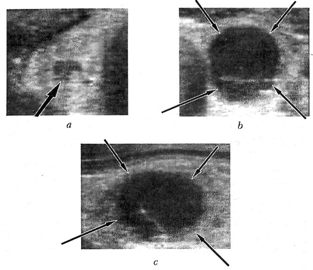
Fig. 2. Patient P., 50 years old. Thyroid gland sonograms.
a — right lobe, cross section, b — left lobe, cross section, c — left lobe, longitudinal section.
In the right lobe (a) round formation: the node (bold arrow) ≈ 8 mm in diameter with heterogeneous inner structure. In the left lobe (b, c) — hypoechogenic
formation: the node, ≈ 23x19x18 mm (marked by arrows)
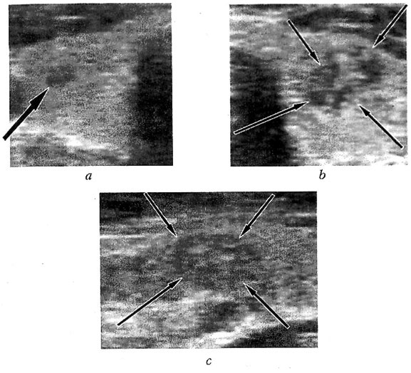
Fig. 3. Patient P., 50 years old. The thyroid gland sonograms in 4.5 months after th

